Brain (cont'd)
The Optic Chiasm
The point where the nasal retinal axons cross, or decussate, is called the optic chiasm. Just above the chiasm is a small grey-matter terminus for certain ganglion cells neurons (...but which?) called the suprachiasmatic nucleus.
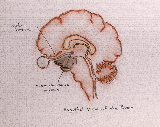
It is a neuronal aggregation common to all vertebrates, initiating the circadian rhythms of the body.
Circadian rhythms are the "regular" physiological changes to which we are subject as we experience the transition from day to night and night to day: changes in core temperature, in heart rate, in hormonal concentrations; the onset of sleep and wakefulness.
In any case, certain ganglion cell axons project to these nuclei in each hemisphere, apparently conveying signals regarding light intensity (information not otherwise emphasized by other nuclei): i.e., information regarding the time of day.
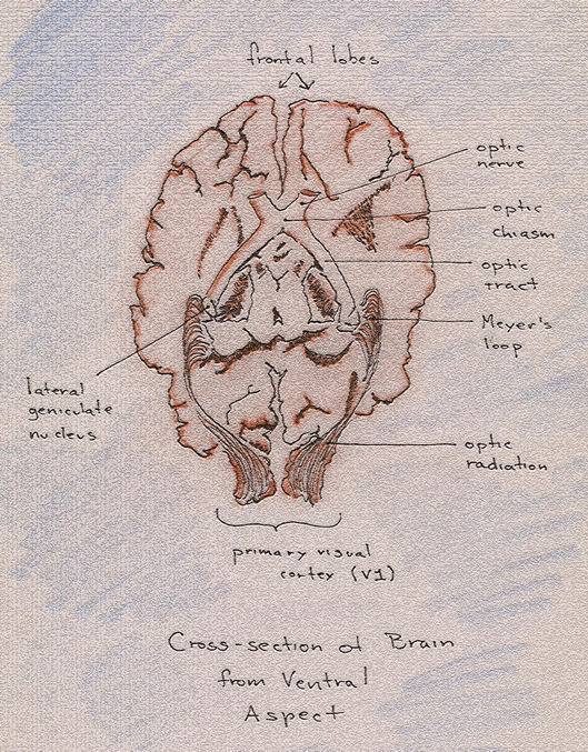
Beyond the optic chiasm, we begin to refer to the optic nerve as the optic tract. Among other things, it allows us to distinguish—admittedly, somewhat grossly—between anterior and medial/posterior locations within the brain—not an insignificant matter when staring at the relative sameness of brain tissue.
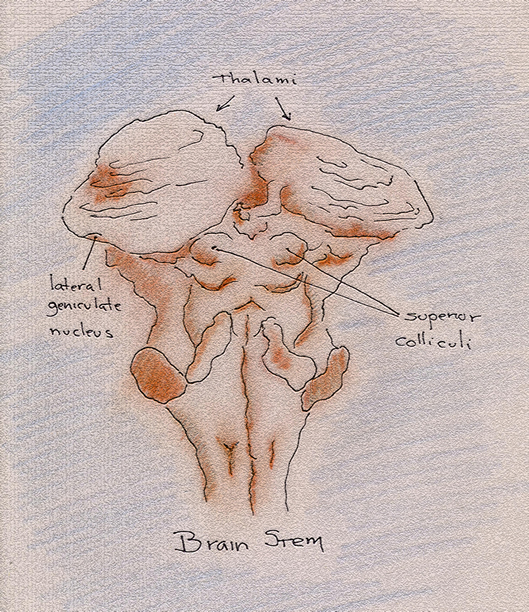
The Superior Colliculi
Beyond the optic chiasm, where the right and left visual field components of one eye have been joined to those of the other, the optic nerve tracts project to the superior colliculi. These are a pair of nuclei that sit atop the midbrain and work to integrate visual and auditory information for the purposes of guiding the head and eyes towards that moment's area of interest in the world without. (In non-mammalian species, these nuclei comprise the tectum, which appears to be the most important visual area in the brains of fish, amphibians and reptiles.)
It is an amazing thing to know that not only has the great tangle of ganglion cell axons projected ipsi- and contra-laterally to join visual fields from one eye and the other but that these axons have also projected themselves against the upper layers of the superior colliculi to form a retinotopic map: i.e., a map whose points correspond to points on the retina arranged in the same relative positions.
The map of the superior colliculi, however, involves a certain bias known as retinotopic magnification. This bias arises from the relative differences in the number of ganglion cell projections from the various parts of the retina. The fovea is the area of highest visual acuity and, as such, projects a larger number of ganglion cell axons to the brain than do peripheral regions of the retina. As a result, retinotopic maps in the superior colliculi and primary visual cortex have disproportionately large areas devoted to the fovea when compared to the actual surface area that it occupies in the retina.
Ganglion cell axons also project to deeper layers within the superior colliculi, which participate in eye and head movement.